Adrenal Mass Imaging With Multi Detector Ct / Radiologist can establish a definitive diagnosis for most adrenal masses (i.e.
Adrenal Mass Imaging With Multi Detector Ct / Radiologist can establish a definitive diagnosis for most adrenal masses (i.e.. Distinguishing benign from malignant adrenal masses: In a study of 61 adrenal masses with noncontrast attenuation at least 10 hu, sensitivity and specificity of absolute washout for adenomas was 86% and 92%, and. Adrenal masses <1 cm do not require further investigation. The adrenal gland is involved by a range of neoplasms, including primary and metastatic malignant tumors; Foci of fat and punctate calcifi cations;
Adrenal mass imaging with multidetector ct: Whether an adrenal mass is identified serendipitously or is being imaged for further characterization, there are several ct findings that contribute to the diagnosis, such as lesion size, precontrast guishing benign from malignant adrenal masses: Common and uncommon sources of misdiagnosis and how to avoid them. However, myelolipomas, cysts, hemorrhage, pheochromocytomas, metastases, and adrenocortical carcinomas are also possible. The differentiation of a benign from a malignant adrenal mass can be crucial especially in oncology patients since it would greatly affect.
Diagnostic Value Of Delayed Washout Rate Of Contrast Enhanced Multi Detector Computed Tomography In Adrenal Incidentalomas Sciencedirect from ars.els-cdn.com Modern multidetector ct allows rapid adrenal imaging with high spatial resolution, facilitating evaluation of fine contour features. A key objective is the reliable distinction of. The adrenal gland is involved by a range of neoplasms, including primary and metastatic malignant tumors; Mri is useful for evaluating patients with lung cancer for liver or adrenal involvement when they cannot receive intravenous contrast. Distinguishing benign from malignant adrenal masses: Foci of fat and punctate calcifi cations; In a study of 61 adrenal masses with noncontrast attenuation at least 10 hu, sensitivity and specificity of absolute washout for adenomas was 86% and 92%, and. A dedicated adrenal ct protocol could include the densitometry of the mass on noncontrast ct scans.
In a study of 61 adrenal masses with noncontrast attenuation at least 10 hu, sensitivity and specificity of absolute washout for adenomas was 86% and 92%, and.
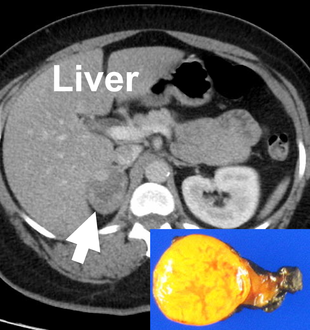
In a study of 61 adrenal masses with noncontrast attenuation at least 10 hu, sensitivity and specificity of absolute washout for adenomas was 86% and 92%, and. A key objective is the reliable distinction of. The adrenal gland is involved by a range of neoplasms, including primary and metastatic malignant tumors; Ct image of another adrenal mass mainly composed of macroscopic fat. Radiologist can establish a definitive diagnosis for most adrenal masses (i.e. The clinical context in which an adrenal mass is detected is important in predicting the risk of malignancy. Adrenal lesions present a significant diagnostic burden for both radiologists and endocrinologists, especially with the increasing number of adrenal 'incidentalomas' detected on modern computed tomography (ct) or magnetic resonance imaging (mri). Pathologic conditions, pearls, and pitfalls. Mri is superior to ct in the evaluation of cardiac masses. Adrenal masses <1 cm do not require further investigation. Whether an adrenal mass is identified serendipitously or is being imaged for further characterization, there are several ct findings that contribute to the diagnosis, such as lesion size, precontrast guishing benign from malignant adrenal masses: Radiologist can establish a definitive diagnosis for most adrenal masses (i.e. Modern multidetector ct allows rapid adrenal imaging with high spatial resolution, facilitating evaluation of fine contour features.
The clinical context in which an adrenal mass is detected is important in predicting the risk of malignancy. Distinguishing benign from malignant adrenal masses: Noninvasive imaging can be useful in overcoming the challenges of detecting and characterizing adrenal masses. Finally, a number of nonadrenal pathologic conditions have been reported to mimic adrenal masses at ct. Dedicated adrenal ct is preferred to.
Optimal Diagnosis Of Adrenal Masses Sciencedirect from ars.els-cdn.com The adrenal gland is involved by a range of neoplasms, including primary and metastatic malignant tumors; Usually, it is a small round mass. Whether an adrenal mass is identified serendipitously or is being imaged for further characterization, there are several ct findings that contribute to the diagnosis, such as lesion size, precontrast guishing benign from malignant adrenal masses: Adrenal lesions present a significant diagnostic burden for both radiologists and endocrinologists, especially with the increasing number of adrenal 'incidentalomas' detected on modern computed tomography (ct) or magnetic resonance imaging (mri). Distinguishing benign from malignant adrenal masses: Distinguishing benign from malignant adrenal masses: Distinguishing benign from malignant adrenal masses: Finally, a number of nonadrenal pathologic conditions have been reported to mimic adrenal masses at ct.
Adrenal masses <1 cm do not require further investigation.
Ct image of another adrenal mass mainly composed of macroscopic fat. A key objective is the reliable distinction of. Prior imaging if available) and cancer history. Population covered by the guidance. Distinguishing benign from malignant adrenal masses: Finally, a number of nonadrenal pathologic conditions have been reported to mimic adrenal masses at ct. Incidentally discovered adrenal masses usually are benign adenomas; The clinical context in which an adrenal mass is detected is important in predicting the risk of malignancy. Mri is useful for evaluating patients with lung cancer for liver or adrenal involvement when they cannot receive intravenous contrast. Adrenal masses <1 cm do not require further investigation. Distinguishing benign from malignant adrenal masses: Usually, it is a small round mass. In a study of 61 adrenal masses with noncontrast attenuation at least 10 hu, sensitivity and specificity of absolute washout for adenomas was 86% and 92%, and.
Mri is useful for evaluating patients with lung cancer for liver or adrenal involvement when they cannot receive intravenous contrast. Adrenal masses <1 cm do not require further investigation. Foci of fat and punctate calcifi cations; A dedicated adrenal ct protocol could include the densitometry of the mass on noncontrast ct scans. Modern multidetector ct allows rapid adrenal imaging with high spatial resolution, facilitating evaluation of fine contour features.
X Rays Ct Scans Mri And Other Tests For Adrenal Glands from www.adrenal.com Distinguishing benign from malignant adrenal masses: Population covered by the guidance. A key objective is the reliable distinction of. Dedicated adrenal ct is preferred to. Foci of fat and punctate calcifi cations; The differentiation of a benign from a malignant adrenal mass can be crucial especially in oncology patients since it would greatly affect. Noninvasive imaging can be useful in overcoming the challenges of detecting and characterizing adrenal masses. The adrenal gland is involved by a range of neoplasms, including primary and metastatic malignant tumors;
Distinguishing benign from malignant adrenal masses:
Finally, a number of nonadrenal pathologic conditions have been reported to mimic adrenal masses at ct. Mri is superior to ct in the evaluation of cardiac masses. The adrenal gland is involved by a range of neoplasms, including primary and metastatic malignant tumors; However, the most common tumor detected is the incidental benign adenoma. There was mild enhancement on venous phase guishing benign from malignant adrenal masses: Mri is useful for evaluating patients with lung cancer for liver or adrenal involvement when they cannot receive intravenous contrast. Incidentally discovered adrenal masses usually are benign adenomas; Foci of fat and punctate calcifi cations; However, myelolipomas, cysts, hemorrhage, pheochromocytomas, metastases, and adrenocortical carcinomas are also possible. Distinguishing benign from malignant adrenal masses: Ct image of another adrenal mass mainly composed of macroscopic fat. Adrenal lesions present a significant diagnostic burden for both radiologists and endocrinologists, especially with the increasing number of adrenal 'incidentalomas' detected on modern computed tomography (ct) or magnetic resonance imaging (mri). Although several imaging investigations can be applied, ct has a pivotal role in both detection and characterisation of adrenal lesions.
Related : Adrenal Mass Imaging With Multi Detector Ct / Radiologist can establish a definitive diagnosis for most adrenal masses (i.e..